6.1. Mammary Gland Development
Mammary gland is the milk-secreting gland in female animals post-partum, although some species produce milk peripartum. The mammary gland is a defining feature of mammals. Mammary gland development commences embryonically and continues post-natal even during lactation. The mammary gland begins developing in utero at approximately 30 d gestation in dairy cattle.
At birth, calves have an udder consisting of basic internal structures, such as small ducts and a mammary fat pad, and fully developed outer structures, such as teats. Before weaning, the rudimentary ducts will form major ducts and the mass of the mammary parenchyma and mammary fat pad increase, although there is little to no lobuloalveolar development.
The study of the mammary gland is important to understand the physiological mechanisms involved during its growth and development, during its functional activity, and to control some pathologies and external factors that can affect its secretory capacity.
Histologically, the mammary gland is a tubule alveolar gland of ectodermal origin, a modified apocrine sweat gland that is found in both sexes but only completes its full development in the female. In the case of the cow, it is called the udder.
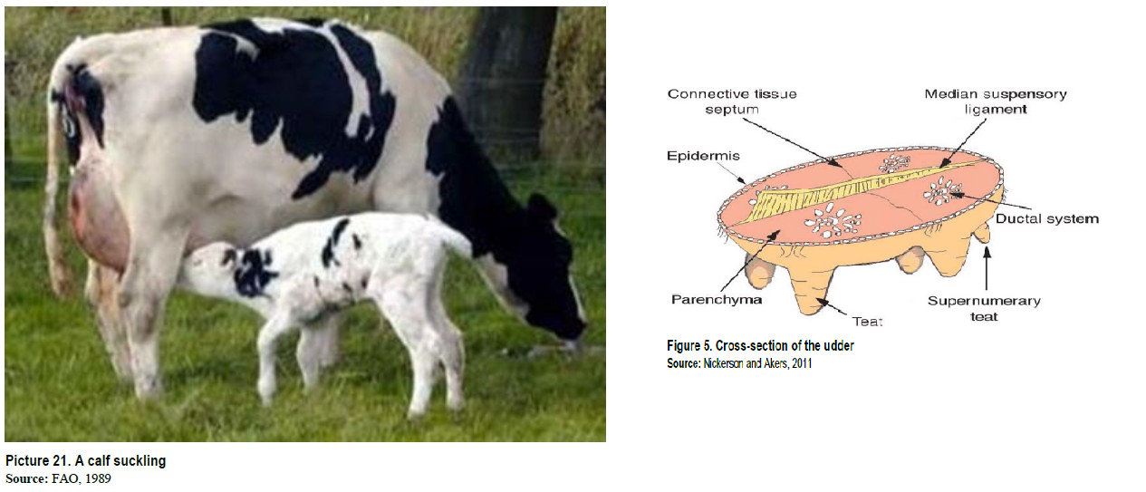
Externally, the udder is a set of four functional glands gathered in a single mass, it has a parallelepiped or saccular shape, and it is located in the inguinal region, suspended by the medial and lateral suspensory ligaments. It is divided into two symmetrical lateral halves by a sagittal furrow called the mammary groove, each of these halves is divided in turn, into two anterior and posterior quarters, although this separation in some cases is not very evident. Each quarter is projected inferiorly by a cylindrical protrusion called the nipple (Figure 5).
The udder is covered with short, smooth, and sparse hair, except on the nipples. The rear quarters are generally of greater capacity than the front quarters. In the specific case of the dairy cow, the udder must be wide, located between the hind legs, and long, extending from the floor of the belly in the umbilical region (anterior insertion) to the perineal region (posterior insertion), also it must have a moderate depth that is determined by the support provided by the firm and elastic suspensory ligaments called the medial and two lateral ligaments.
The internal structure of the udder is composed of secretory parenchyma and a connective tissue stroma.